Testing for Gout
Learn about the lab and imaging tests used to diagnose and track gout.
By Mary Anne Dunkin | June 10, 2022
Sometimes gout can be diagnosed based only on symptoms: intense, painful swelling in a single joint — often in the big toe — that subsides, only to show up again in the same or a different joint. In other cases, it may be more difficult to pinpoint the cause of your joint pain, so a doctor may order lab or imaging tests to confirm or reject a gout diagnosis, or to make sure it’s not a different condition.
Once a diagnosis is confirmed and you’re receiving treatment, you may need to undergo further tests to monitor the disease progression or how well a treatment is working.
Here are some of the tests you may experience when you have, or think you have, gout.
Diagnostic Lab Tests
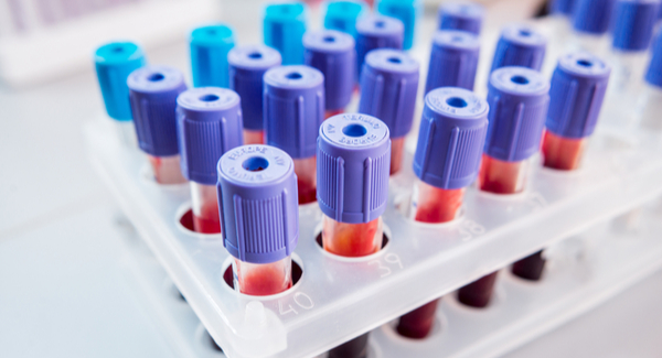
Different lab tests for gout examine either the patient’s blood or the fluid in their affected joint. The most common tests are for:
Uric acid. Gout occurs when uric acid, a bodily waste product, builds up in the bloodstream and forms as crystals in the joints. A blood test that shows high levels of uric acid could help confirm a diagnosis of gout. However, not all people with gout have high levels of uric acid in the blood and not all people with high uric acid levels have gout.
Joint fluid analysis. In this test your doctor will insert a needle into the affected joint and draw out some synovial fluid to check for the presence of uric acid crystals. This is a more accurate way to confirm a gout diagnosis.
Diagnostic Imaging Tests
Imaging tests may help rule out other causes of joint pain and inflammation as well as to identify crystals or joint changes indicative of gout. Imaging tests used to diagnose gout may include:
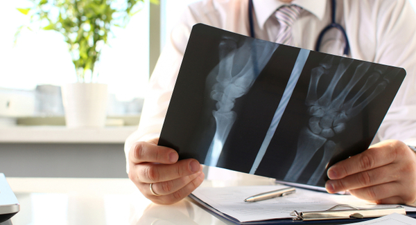
X-ray. Joint damage from gout may not be visible until years after the onset of gout symptoms, so the primary benefit of X-rays in diagnosing gout is to rule out other potential causes of joint pain.
Ultrasound. Ultrasound, or sonography, uses sound waves to create pictures of structures inside the body. It may be used to detect urate crystals in the joints or deposits of uric acid crystals that form hard, visible lumps in or near the joints, called tophi.
Dual-energy computerized tomography (DECT). A test that combines X-ray images from many different angles, DECT may be used to see uric acid crystals in the joints.
Monitoring Lab Tests
Because treatment for gout is targeted at lowering levels of uric acid in the blood to a target of 6 milligrams per deciliter (6 mg/dL) or lower, blood tests to check uric acid levels are performed periodically to monitor the effectiveness of treatment. Your doctor will use the results of this test to adjust your medication and other treatments.
Other tests your doctor may order include:
Creatinine. This test measures the levels of creatinine, a waste product made by your muscles, in a sample of blood or urine. Abnormal levels could be a sign of kidney disease.
Blood Urea Nitrogen (BUN). Often performed in conjunction with the creatinine test, this test checks for blood levels of urea nitrogen, a bodily waste product normally excreted through the kidneys. A high level could mean kidneys are not working properly.
Urinalysis. Your doctor may order check for high levels of uric acid in your urine, which could indicate a risk for kidney stones.
Monitoring Imaging Tests
If not well controlled, gout can cause damage to the joints. Your doctor may order one of the following imaging tests to check for joint damage.
- X-ray
- Ultrasound
- Magnetic resonance imaging (MRI) to produces 3D images of joints or other tissues.
- Computed tomography (CT) scan, which combines a series of X-ray images to create cross-sectional images of parts of the body.
Stay in the Know. Live in the Yes.
Get involved with the arthritis community. Tell us a little about yourself and, based on your interests, you’ll receive emails packed with the latest information and resources to live your best life and connect with others.


